3D rotational angiography (AKA flat-panel volume CT or cone-beam CT) is a powerful tool in the assessment of vascular anatomy and aneurysm morphology. The technique works by spinning the C-arm around the patient, taking 10s to 100s of images while contrast is injected in the vasculature. Reconstructions can then be generated using software.
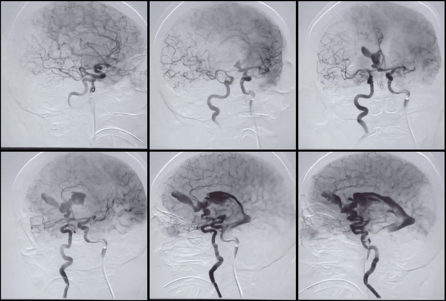
This is what you DON’T want to see during your 3D rotation angiogram (via NEJM):

Some lessons to consider for this angiogram… The intermediate catheter is placed almost in the cavernous segment of the internal carotid artery, which is too high for a 3D rotational angiogram in a recently ruptured aneurysm case. The rate and volume were too high for this injection (as indicated by reflux into the contralateral cervical ICA and entire contralateral intracranial circulation. There is also spasm around the guide catheter which increases the pressure of the injection. It’s better to conduct these angiograms through a diagnostic catheter from the proximal ICA or even the CCA to reduce the risk of catastrophic hemorrhage.
