The vertebral arteries are paired arteries (left and right) that typically branch from the subclavian arteries respectively and supply the posterior fossa and the occipital lobes. In approximately 6% of people, the vertebral artery can arise directly from the aortic arch (typically on the left).
The vertebral artery is divided in 4 segments:
- “V1: pre-foraminal segment
- origin to the transverse foramen of C6
- V2: foraminal segment
- from the transverse foramen of C6 to the transverse foramen of C2
- V3: atlantic, extradural or extraspinal segment
- starts from C2, where the artery loops and turns lateral to ascend into the transverse foramen
- continues through C1 to pierce the dura
- V4: intradural or intracranial segment
- from the dura at the lateral edge of the posterior atlanto-occipital membrane to their confluence on the medulla to form the basilar artery”
Of course, the vertebral artery is intimately connecting to the cervical muscles, spine, and carotid arteries.
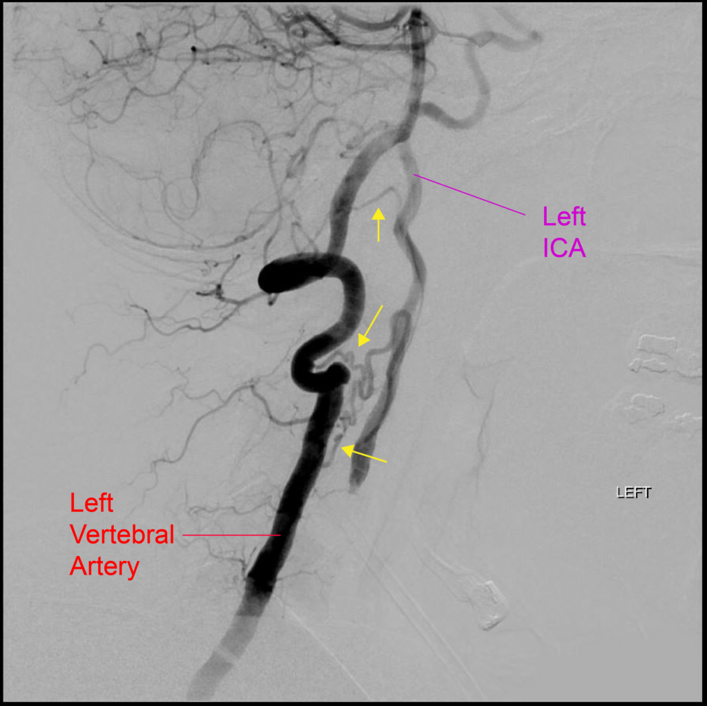
Here is an interesting example of collateralization from the left vertebral artery eventually to the left ICA. In this case, the left ICA had been chronically occluded and these collaterals developed over time to provide support to the right anterior circulation.

As you can see, there are least three separate branches (yellow arrows) arising from the left vertebral artery that eventually supply the left ICA. The superior branch is likely a collateral traveling through the odontoid arcade back to the ascending pharyngeal artery (which then feeds retrograde into the left ICA). The lower two branches are likely hypertrophied C2 and C3 segmental arterio-arterial collaterals to the musculospinal branch of the ascending pharyngeal artery, which then also feeds retrogradely back into the left ICA.
