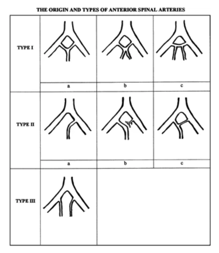
The anterior spinal artery (ASA) is a small yet critical artery that supplies the anterior surface of the spinal cord (e.g. motor tracts). It typically arises from the V4 segment of the vertebral arteries (near the medulla) and co-joins to form a single artery that overlies the anterior longitudinal fissure of the spinal cord. It give off a sulcal artery at most spinal levels, which enters the anterior median fissure.
The outer diameter of the ASA ranges from 0.34 to 1.02 mm (mean 0.59 mm) (reference).

There is tremendous variability in the origins and types of anterior spinal arteries. The classical “textbook” ASA, called Type Ia, was not observed in one small series of nine cadaver samples (reference).
Along the course of the anterior spinal artery, it is reinforced at several levels:
~C3: Radiculomedullary artery from the vertebral artery
~C6/7: Ascending cervical artery
~T3-T7: Posterior intercostal arteries
~T8-conus: The Artery of Adamkiewicz
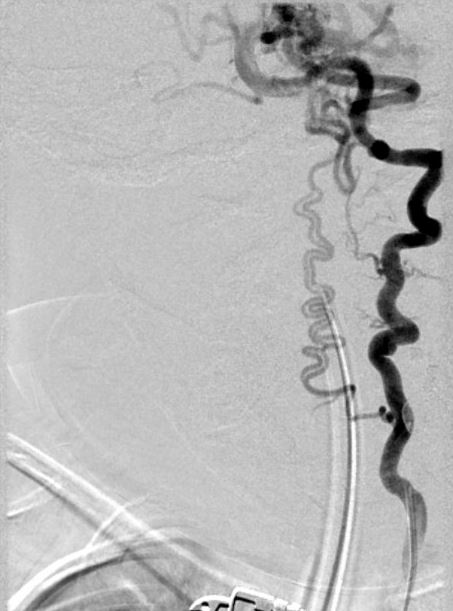
Here is an example of a duplicated anterior spinal artery in the case of a young girl with a posterior fossa plexiform arteriovenous malformation. The artery arises from a cervical radiculomedullary branch of the left vertebral artery and traverses superiorly. In this case, the AVM is demanding flow from the duplicated anterior spinal arteries.

It is likely that the enhanced hemodynamic requirements of the AVM resulted in hypertrophy of an embryologic duplicated anterior spinal artery.
