This is a patient in his early 60s who presented to the hospital as a code stroke activation after he was found on the ground unresponsive. Son reported he is a heavy drinker and heavy smoker. Initial NIHSS 9 for right facial droop, right hemianopia to L eye, expressive aphasia and dysarthria. However, no weakness to any extremity but decreased sensation on the right side.
He was taken for CT Imaging which demonstrated:
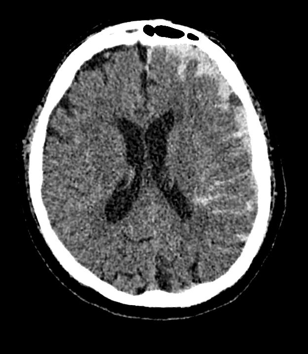
Left parafalcine subdural hematomas. A right anterior frontal lobe parasagittal subarachnoid hemorrhage,
contusion, versus subdural hematoma.
CTA demonstrated possible hypervascular lesion versus active contrast extravasation at the dural region of the left frontal lobe, with a consideration for possible dural AVF or AVM or Aneurysm.
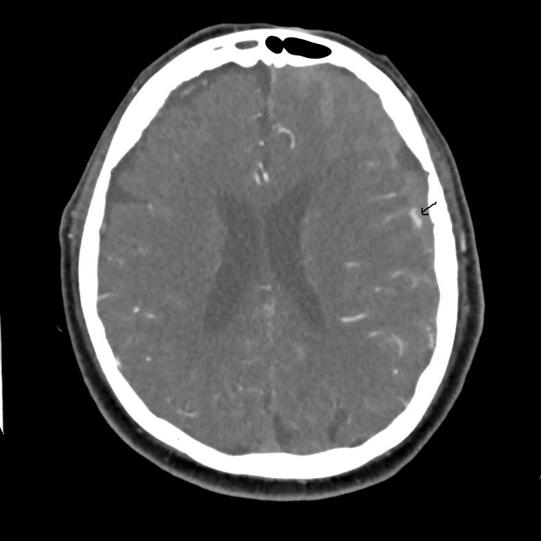
Patient was then taken to IR for a diagnostic cerebral angiogram.
This prompted at stat CT on the table which demonstrated the following:
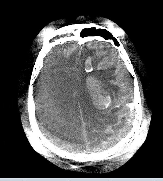
Neurosurgery was immediately alerted and the patient was taken for stat left craniotomy for evacuation of the acute intracerebral hemorrhage and insertion of subdural drain.
Unfortunately, he did not survive this catastrophic event.